Ultrasound Physics
This line of work focuses on quantitative imaging and image reconstruction in diagnostic and interventional clinical applications, with research extending to areas such as tissue biomechanical characterization, inverse problems in imaging, and signal processing. Novel imaging contrast techniques, especially based on ultrasound physics, is a major interest in the group.
Below some of our projects related to this field of activity:
Speed of Sound Imaging
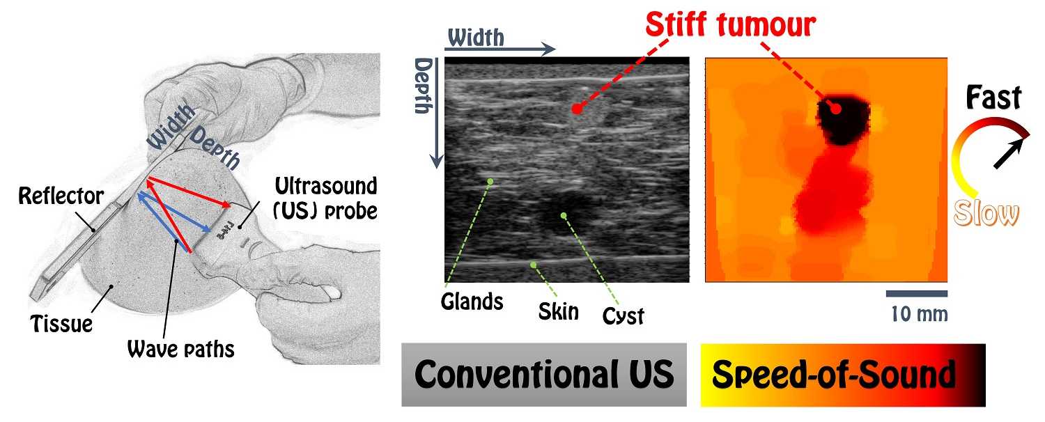
A novel ultrasound imaging contrast modality, speed-of-sound, developed in CAiM received the 2016 ETH Spark Award (for the most promising innovation of the year) and the 2017 Swiss Venture Award (for the best idea). This contrast marker has so far been tested in clinical studies on the breast and on muscles, e.g. for the quantification of breast density and muscular degeneration, as well as for differential diagnosis of breast cancer.
Elastography: Imaging Soft-Tissue Biomechanical Characteristics
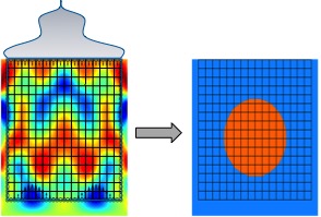
Tissue elasticity has been used in medicine as an indication of tissue anomalies for centuries. Palpation of breast and prostate are still the most common clinical procedures in cancer screening for those corresponding anatomical structures. Elastography is an emerging technique for imaging tissue viscoelasticity. This project aims to address some of the pending issues in generating ultrasound elastography images, while also investigating novel acquisition and analysis techniques for advanced imaging applications.
Elastography is an emerging technique for imaging tissue viscoelasticity from internal tissue displacements observed often in magnetic resonance or ultrasound time sequences. Elastography based on ultrasound is particularly important, as ultrasound is real-time, cost-effective, non-ionizing, and thus a widely-used imaging modality. Elastography has great potential in diagnosis and treatment planning because pathologies, e.g. tumors and cirrhosis, are often stiffer than their surrounding tissue. It can furthermore be beneficial as an indication of tissue composition for robust characterization and segmentation of anatomical structures with different mechanical properties, e.g. for arterial plaque characterization. Despite all these benefits, elastography is still not fully exploited in clinics. Several different methodological approaches proposed in this field are still not fully mature and there is a clear need for robust methods for imaging tissue elasticity in an accurate, fast, and reproducible way. This project aims to address some of the pending issues in generating ultrasound elastography images, while also investigating novel acquisition and analysis techniques for advanced imaging applications.
Different imaging techniques and modalities provide additional information to physicians for better diagnosis and screening, and tissue elasticity is one such information that clinicians can exploit in their decision making. Results of our research will improve current clinical practices by helping to bring tissue elasticity imaging into the arsenal of everyday medicine.